Home
Uncategories
Loculated Pleural Effusion X Ray / Clinical Vignette 27 Miphidic : Other signs on the chest radiograph may suggest a malignant cause for the effusion.
Loculated Pleural Effusion X Ray / Clinical Vignette 27 Miphidic : Other signs on the chest radiograph may suggest a malignant cause for the effusion.
Loculated Pleural Effusion X Ray / Clinical Vignette 27 Miphidic : Other signs on the chest radiograph may suggest a malignant cause for the effusion.. Loculated pleural effusion x ray / diagnostic utility and clinical application of imaging for pleural space infections chest / loculated pleural effusion masquerading as mediastinal tumour had been reported but pleural effusion that conformed to the contour of a lung lobe is rare. Pocus demonstrated a large right sided loculated pleural effusion with associated septations and surrounding consolidation suggestive of a parapneumonic effusion. Surgical thoracostomy tube placement and radiologically guided catheter drainage are standard therapy for loculated pleural fluid collections. The most prominent finding of this scan is a loculated pocket of pleural fluid that does not otherwise appear to extend inferiorly between the right lower lobe and the diaphragm. Large effusions, even if loculated, can cause tracheal.
Dome shaped opacity projecting into the lung noted tracking along the cp angle and lateral chest wall suggestive of loculated pleural effusion, however the possibility of empyema can not be ruled out completely. Loculated pleural effusion x ray / disease of the pleura radiology key : A right thoracentesis was performed, and on seeing the biochemistry results, the left side was also punctured. The right pe was larger and loculated (by ultrasound). Normally, the space between the visceral pleura and the parietal pleura cannot be seen.
Xmlinkhub from e-kcj.org The patient may have unrelated symptoms due to the disease or condition that has caused the effusion.symptoms of pleural effusion include: This is for educational purpose. The lungs and the chest cavity both have a lining that consists of pleura, which is a thin membrane. Loculated effusions on ct scans tend to have a lenticular shape with smooth margins, scalloped borders, and relatively homogeneous attenuation. The presence of pleural fluid was not related to infarction. The most prominent finding of this scan is a loculated pocket of pleural fluid that does not otherwise appear to extend inferiorly between the right lower lobe and the diaphragm. Ct shows heterogeneously enhancing mass lesion left hemithorax (arrowhead) causing mediastinal displacement to the right 94. We present a unique case in which a patient presented to the ed in respiratory distress.
The presence of pleural fluid was not related to infarction.
Blunting of the lateral costophrenic angle usually requires about 175 ml but may take as much as 500 ml. Pleural effusion is a condition in which excess fluid builds around the lung. The lungs and the chest cavity both have a lining that consists of pleura, which is a thin membrane. The excess accumulation of fluid can usually be seen on these images. In patients with loculated pleural fluid the diagnosis of pe had been delayed for a mean of 12.2 days after symptoms developed. Thoracoscopy was considered dangerous because of the patient's extreme fragility and hemodynamic instability. The patient may have unrelated symptoms due to the disease or condition that has caused the effusion.symptoms of pleural effusion include: Other signs on the chest radiograph may suggest a malignant cause for the effusion. Dome shaped opacity projecting into the lung noted tracking along the cp angle and lateral chest wall suggestive of loculated pleural effusion, however the possibility of empyema can not be ruled out completely. Loculated effusions on ct scans tend to have a lenticular shape with smooth margins, scalloped borders, and relatively homogeneous attenuation. Prior chest radiographs indicating that the blunting is a new finding also provide a good indicator of pleural effusion. Pleural effusion with nodes or mass or lytic bone lesions: The pleura and pleural spaces are only visible when abnormal.
This is for educational purpose. A pleural effusion infiltrates the space between these layers. Other signs on the chest radiograph may suggest a malignant cause for the effusion. Read more z2jeetendra medical student follow 10 comments 73 likes. In the context of a large effusion, mediastinal shift toward the side of the effusion should alert the clinician to the possibility of bronchial obstruction, which may.
Pleural Effusion X Ray Findings from image.slidesharecdn.com Treatment may fail if the catheter is not placed optimally within the loculation or if the fluid is hemorrhagic or fibrinous. Pleural effusions are characterized on ct by attenuation values between those of water (0 hounsfield units hu) and soft tissue (approximately 100 hu), typically in the order of 10 to 20 hu. Pleural effusion with a missing breast suggesting resection for cancer : Dome shaped opacity projecting into the lung noted tracking along the cp angle and lateral chest wall suggestive of loculated pleural effusion, however the possibility of empyema can not be ruled out completely. The patient may have unrelated symptoms due to the disease or condition that has caused the effusion.symptoms of pleural effusion include: These can show if the. Normally, the space between the visceral pleura and the parietal pleura cannot be seen. We studied the value of transca …
Loculated effusions are difficult to confirm with chest radiograph, but ultrasound, computed tomography (ct), and even magnetic resonance imaging (mri) may be used to verify a localized collection of pleural fluid.
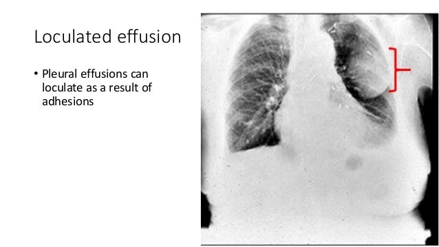
Pleural effusions are characterized on ct by attenuation values between those of water (0 hounsfield units hu) and soft tissue (approximately 100 hu), typically in the order of 10 to 20 hu. The excess accumulation of fluid can usually be seen on these images. Loculated effusion • pleural effusions can loculate as a result of adhesions 17. We present a unique case in which a patient presented to the ed in respiratory distress. Pleural effusion with nodes or mass or lytic bone lesions: Loculated pleural effusion x ray / diagnostic utility and clinical application of imaging for pleural space infections chest / loculated pleural effusion masquerading as mediastinal tumour had been reported but pleural effusion that conformed to the contour of a lung lobe is rare. Pleural effusions can also form when there is transport of peritoneal fluid from the abdominal cavity through the diaphragm or via lymphatics from. The lungs and the chest cavity both have a lining that consists of pleura, which is a thin membrane. Solitary peripheral masses are more likely peripheral subpleural pulmonary masses while pleural masses such as metastases and mesothelioma are most often multiple. Large effusions, even if loculated, can cause tracheal. The effusion appears to have a mildly echogenic, 'speckled' appearance, which is suggestive of exudative effusion. A pleural effusion infiltrates the space between these layers. This is for educational purpose.
A lateral decubitus projection is most sensitive, able to identify even a small amount of fluid. Ct shows heterogeneously enhancing mass lesion left hemithorax (arrowhead) causing mediastinal displacement to the right 94. Because the pleural effusion has a density similar to water, it can be seen on radiographs. These can show if the. Thoracoscopy was considered dangerous because of the patient's extreme fragility and hemodynamic instability.
Chest X Ray Shows Cardiomegaly Enlarged Heart With Infiltration Stock Photo Picture And Royalty Free Image Image 81339182 from previews.123rf.com Malignant fibrous tumor of pleura : Prior chest radiographs indicating that the blunting is a new finding also provide a good indicator of pleural effusion. Read more z2jeetendra medical student follow 10 comments 73 likes. Dome shaped opacity projecting into the lung noted tracking along the cp angle and lateral chest wall suggestive of loculated pleural effusion, however the possibility of empyema can not be ruled out completely. The effusion appears to have a mildly echogenic, 'speckled' appearance, which is suggestive of exudative effusion. Pleural effusions are characterized on ct by attenuation values between those of water (0 hounsfield units hu) and soft tissue (approximately 100 hu), typically in the order of 10 to 20 hu. The right pe was larger and loculated (by ultrasound). In patients with mediastinal lymphoma.
Malignant fibrous tumor of pleura :
In patients with loculated pleural fluid the diagnosis of pe had been delayed for a mean of 12.2 days after symptoms developed. Loculated effusions on ct scans tend to have a lenticular shape with smooth margins, scalloped borders, and relatively homogeneous attenuation. Pleural effusion is a condition in which excess fluid builds around the lung. The effusion appears to have a mildly echogenic, 'speckled' appearance, which is suggestive of exudative effusion. If you miss a tension pneumothorax you risk your patient's. Chest radiographs are the most commonly used examination to assess for the presence of a pleural effusion; We present a unique case in which a patient presented to the ed in respiratory distress. Because the pleural effusion has a density similar to water, it can be seen on radiographs. Loculated pleural effusion x ray / disease of the pleura radiology key : We studied the value of transca … The patient may have unrelated symptoms due to the disease or condition that has caused the effusion.symptoms of pleural effusion include: The pleura and pleural spaces are only visible when abnormal. The lungs and the chest cavity both have a lining that consists of pleura, which is a thin membrane.
If you miss a tension pneumothorax you risk your patient's loculated pleural effusion. Solitary peripheral masses are more likely peripheral subpleural pulmonary masses while pleural masses such as metastases and mesothelioma are most often multiple.


0 Comments:
Posting Komentar